
3D Imaging – CBCT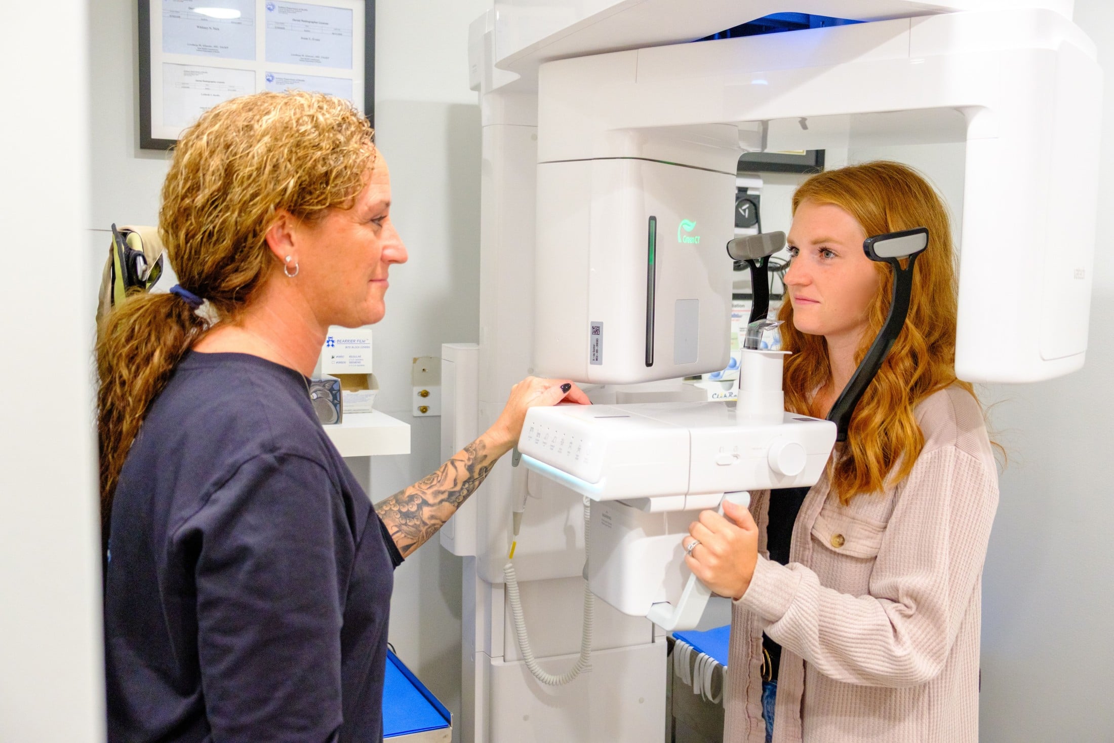
Cone Beam Computed Tomography (CBCT)
Over 35 years of his experience in dentistry, Dr. Burns states one of the most important developments has been the ability to perform an in-office 3-dimensional radiograph.
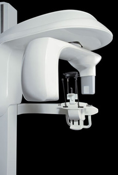
Dr. Burns utilizes the VaTech, Green X, CBCT machine, the most recent and advanced unit in the VaTech CBCT lineup. Dr. Burns has the benefit of seeing many different types of CBCT images in the course of his practice, and he feels that the Green X has one of the clearest images on the market.
The Green X system provides Dr. Burns state-of-the-art technology to help diagnose and treat his patients with unprecedented confidence.
Procedures Enhanced by the Use of CBCT Imaging:
- Implant Placement: CBCT imaging has revolutionized dental implant placement. While in rare instances, it is not absolutely mandatory to have a CBCT scan, having the image aids in diagnosis, planning, and patient safety. It also marries with the X-Guide Dynamic Guidance System to improve implant placement and success. Dr. Burns feels so strongly about the use of CBCT and the X-Guide that he will not place an implant without them.
- Tooth Extractions: At times certain anatomic structures (maxillary sinus, inferior alveolar nerve, etc.) are in close proximity to the tooth to be extracted. The CBCT scan provides the exact location in the 3D space to determine the truest level of risk to these structures and avenues to avoid damage.
- Impacted Tooth Exposure: Because the tooth is impacted, its truest location must be determined in order to plan the best route of exposure. The CBCT scan provides this critical view which aids in guiding Dr. Burns to exposing the tooth for ongoing orthodontic treatment.
- Pathology: The CBCT scan can be a vital tool in diagnosing a lesion that is below the surface of tissue and possibly encased in the jawbone. Once diagnosed, the treatment plan is developed utilizing the CBCT scan of the critical structures. surrounding the pathology.
- Bone Grafting Procedures: The CBCT scan demonstrates the entirety of a bony defect and its effect on determining ideal implant location. Once the graft has healed, further CBCT scans confirm successful bony growth which allows the implant placement to proceed.
- Sinus Lift with Grafting: Prior to a sinus lift, the CBCT scan provides a detailed view of the maxillary sinus, including an anatomy that might complicate a sinus lift and the initial health of the sinus. After the healing period, a secondary CBCT scan confirms successful sinus lift with improved bone in the treatment zone before implant placement.